by Dr. Jeremy Schmoe, DC, DACNB
Many of our patients seek help, hope, and answers for lingering post-concussion symptoms. These injuries may have occurred during sporting events, car accidents, falls, blast injuries or various other modes of trauma. Not only do these injuries lead to chronic dizziness, vertigo, visual symptoms, headaches, and balance issues, but they also affect limbic regions in the brain causing anxiety, depression, startle responses/fear and apathy.
In this article, I am going to touch on some aspects that I see every day in our clinic, and hopefully make some sense of these complex post-concussion symptoms.
It is not uncommon for the patients that I see to have been told by other providers that their sensations of dizziness and vertigo are from anxiety and depression. They are also told that their exam was completely normal, and there is no sign of BPPV. However, people that have experienced a brain injury have multiple sensory hubs and circuits that are affected. This means that it isn’t as simple as stating that the person has BPPV or peripheral vestibular disorder. It is the complex integration of the sensory hubs and circuits that are affected with TBI.
Furthermore, your balance may even be normal when we do a platform posture analysis, even though you are extremely dizzy throughout the day mostly noted with complex visual environments. After visual stimulation you may feel lightheaded, off balance, stiffness in your cervical spine, head pressure, shaky on the inside and/or anxiety. Your energy levels seem to tank out of nowhere, leading to extreme fatigue and brain fog. You need to eat in order to keep your blood sugar steady—or else you know its is a downward spiral that there is no coming back from. All of these scenarios could be due to the fact that you are experiencing visual vertigo.
Anxiety kicks in at night due to the stimulation you experienced during the day. Now you are experiencing restless and sleepless nights. People that you talk to suggest that you work out because they heard that “working out is great for concussion recovery.” In your mind, you are thinking, “Yeah, right, there is no way I am going to be able to muster up enough energy to do anything productive. Let alone if my heart rate increases there is no telling when it will come back down. Do these advice-givers realize that any sort of activity will cause me to not be able to sleep— and the anxiety kicks in more.”
The anxiety about the sleepless nights and overstimulation keeps piling up until finally you crash. It might be 3–4 days later before you are able to get a good night of sleep. This cycle perpetuates for weeks, months, and sometimes years.
You are thinking:
- When is this going to end? Is it ever going to end?
- Who is going to help me? I have seen everyone, and they say I am fine. My family must think I am going insane. How much longer are they going to put up with me?
- How am I ever going to be able to go to college?
- How can I can I take care of my children?
The list goes on and on in your mind until finally your system crashes, leaving you completely in a fog.
These are all common scenarios I hear every day at The Functional Neurology Center where we specialize in post-concussion rehab. And I have had to learn all this from compiling information from multiple specialties, making clinical observations—and treating a lot of suffering patients. For the most part, training for treating concussions is in post-graduate education.
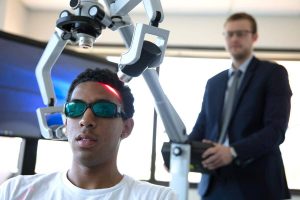
Okay, back to the neurology! Many of these patients are suffering from visually induced vertigo post concussion. Examination would be simpler if only one type of force occurred during trauma; however, multiple different types of forces can occur. Some common ones are linear translation with acceleration and rotation leading to torsion. These forces lead to changes in different sensory-motor hubs and circuits of the brain. Due to the complexity of the visual system and its use of all regions of the brain, you will have visually mediated reflexes affected with almost all TBI’s.
Clinically you may see changes in fixation, pursuits, saccades, OKN reflexes, along with combined head-eye motion. These patients may develop vertiginous symptoms when exposed to visually challenging environments. There are changes in the central projections from the cerebellum into the vestibular nuclei, NPH, oculomotor nuclei, superior colliculus and basal ganglia. Any inaccurate feedback from the eye muscles themselves can cause an unstable visual perception, which does not match with what the feedback from your muscles are telling you. This can even be from a muscle in lower extremity, upper extremity, lumbar paraspinal muscles, thoracic paraspinal muscles, or even your cervical-occipital region. Any of which lead to a sensory error with an impaired ability to have appropriative top-down corollary discharge and efferent (directing outward) copy mechanisms in the cerebellum.
Next thing you know a little bird flies into your field of view, which you perceive as a postural threat. This is because your eyes were not in the center of the globe—potentially due to the square wave jerk you had from loss of superior collicular activation of Omnipause neurons. Omnipause neurons inhibit saccadic burst neurons. Due to the loss of inhibition from the omnipause neurons, you may be experiencing saccadic intrusions, and this affects your ability to know where your head and neck are in space. You can develop cervicogenic headaches, changes in feedback from muscle spindles in the cervical spine that further aggravates the situation.
Your head/neck may feel like it’s going to explode after a trivial visual stimulus.
- If your head feels like it is full with pressure exploding from the inside out, there is usually underlying dysautonomia.
- If your head feels like it is being compressed from the outside in, there is usually a biomechanics cause.
Think about the scenario above and image how anxiety-provoking it would be if that were happening to you all day everyday. The cycle of pain, anxiety, fatigue, dizziness, over-stimulation and other symptoms could continue until you find the right provider who can make sense of what is going on with you.
Not only are the central vestibular connections affected, but also the integration between numerous other circuits. These circuits are the cervical spine afferents, vestibular nuclei, vestibular autonomic, spino-oculomotor connections, trigeminal-oculomotor proprioceptive feedback, vagal-autonomic, brain-gut connections and higher order processing above the thalamus.
When you really break it down, the brainstem connections that go up into the cortex and also down into the cord become affected. The midline regions of the cerebellum and the integration of your spinal stability become dysfunctional as well. This leads to instability in your midline musculature that allows you to stabilize your body as you move your center of pressure. All of this contributes to your symptoms of headaches, neck pain, spinal instability, stiffness, gait changes, head pressure, dizziness, vertigo and light-headed sensations. Don’t forget that many of the limbic regions of the brain are midline, and also get affected. The midline regions of your brain got rung out like a wet towel. You look fine on the outside, but on the inside the major hubs are not communicating with each other. This means that you are unable to navigate your world effectively, along with being unaware of where your body is in space. This leads to difficulty performing tasks such as running, walking, and even breathing!
One mechanism that I see affected in the vast majority of my post-concussion patients is breathing. The breathing mechanics become dysfunctional from changes in central pattern generators in the brainstem. The centers that reflexively allow you to expand your ribs and get oxygen can be injured in TBI. Your brain needs oxygen in order to function optimally and to heal. This could be one reason that people chose to go to clinics with hyperbaric oxygen to help them recover.
A lot of the rehab we perform at my clinic is around getting optimal autonomic function, improved breathing mechanics and oxygenation. The reason behind this may be attributable to the fact that the region of the brainstem that allows you to know where you are in space is right next to the autonomic region in the pontomedullary reticular formation.
For example, if you develop hypoxia to your cerebellum, you could experience down-beating nystagmus, increased gain of your VOR, or tonic deviations of the eyes upward from vestibular disinhibition. This means that your cerebellum and its purkinje neurons are not able to modulate the vestibular nuclei, which leads to centrally mediated vestibular dysfunction from dysautonomia caused by your brain injury.
Many of the patients we see develop POTS and dysautonomia with visual/vestibular symptoms post concussion. These types of issues do not always fit into the main vestibular dysfunctions like BPPV, Meniere’s, Vestibular Migraine, Vestibular Neuritis, Phobic Postural Vertigo or MdDs. Traumatic Brain Injury-induced vestibular, visual and proprioceptive dysfunction is quite complex and doesn’t follow textbook protocol as each person is biochemically unique and may present with previous underlying co-morbidities. This is why in addition to neuro-rehabilitation, our clinic believes it is important to address your nutrition and run comprehensive lab work. We also address underlying brain-based inflammation and microglia priming with non-invasive low-level cranial laser. By addressing inflammation and providing the brain with optimal nutrition and fatty acids, this allows our neuro-rehabilitation to build plasticity.
The autonomic system may become dysregulated from trauma. If this occurs, one may see a decrease in venous return along with pooling of blood in the pelvis and legs due to improper vasoconstriction and dilation. This may lead to the feeling of light-headedness, syncope, and/or brain fog.
Due to the presentation of these symptoms mentioned above, this is why we focus on working out the legs in our patients with post-concussion symptoms. We like to create an environment that artificially activates the patient’s leg muscles while they either sit or lay. We utilize Arpwave Neuro stimulation techniques along with isometric contractions to provide feedback to the brain. Once the patient becomes more stable, we move into more complex motion and eccentric/concentric leg exercises.
In our clinic we commonly assess the VOR reflex, which occurs when you move someone’s head one way and their eyes move back equal and opposite to have a gain of 1. There can be increased and/or decreased gain in patients. Most of the head movements we experience in every day life are fast and unpredictable and that is why clinically subtle VOR dysfunction can be missed. Everything from a clinical standpoint goes back to improving VOR gain, gaze stability and stabilizing autonomics.
To know where you are in space requires constant adjustments made by the body to gravitational load. There is proprioceptive feedback from external ocular muscles (EOMs). The position sense of the eyes in the orbit are used to help calculate the position of the target in your world with respect to your body. When these systems are affected, you can develop positional errors in the body such as a neck position error. These errors of the neck commonly are in the “yaw, pitch and roll” planes which are coordinated with the vestibular system centrally. Any minuscule error that is maintained from injury to the central hubs may affect the sensation of depth, position, or saccadic (eye movement) accuracy. One example is injury to the cerebellum. This can lead to sensory mismatch with an impaired ability to adapt and heal.
We can improve function in our patients with post-concussion symptoms by performing specific combinations of head and eye movements. Many of our patients have performed visual exercises prior to seeing us with success, but are still experiencing lingering symptoms. For patients that are unable to tolerate occulomotor and/or vestibular rehab, our clinic utilizes non-invasive nerve stimulation. This may make the performance of visual exercises easier.
Many patients post brain injury develop Convergence Insufficiency (CI). This can lead to symptoms such as painful eye movements, headaches, or even attentional issues. There most likely are contributions from the superior rectus during convergence in patients with convergence insufficiency. The superior rectus can provide adduction and incyclotorsion of the eyes to make up for impaired ability of the medial rectus to perform its duty. There will be more sympathetic tone needed to allow these muscles to do this job. There could be a need to use the Superior Rectus to maintain binocular focusing for clear images on gaze fixation. The EOMs are known to be fatigue resistant, but obviously so many patients experience pain, tightness and spasm post concussion that there must be fatigue. This fatigue develops with their high metabolic demand of static contraction of muscles being used in their secondary and tertiary actions to make up for convergence insufficiency.
If a patient has developed a spastic contraction of an ocular motor muscle in intorsion, their brain may adapt with a roll-plane deviation of the head or skew deviation of the eyes leading to depth-field contamination of OKN response. This can lead to anxiety, visual motion illusion, dysautonomia and/or even headaches. These types of patients have a difficult time going into Target, the grocery store, or even malls. When driving in busy traffic these patients may experience the sensation that cars are coming into their lane.
The mismatch of the incoming proprioceptive information from the EOM’s may not match perfectly with the other senses such as vestibular ocular and spino-ocular from impaired cerebellar coordination. This could lead to an increased sympathetic response that further leads to increased adrenergic tone and even dysautonomia. The EOM’s become spastic in their ability to move.
The patient could have these issues:
- convergence insufficiency, spasm on convergence,
- impaired accommodation,
- gaze instability,
- changes in pursuits,
- saccadic accuracy,
- inability to tolerate motion,
- cerebellar dysfunction,
- cervical spinal dysfunction,
- central and peripheral vestibular dysfunction with nystagmus.
All of this leads up to visual illusion with the patients unable to tell you what it looks or feels like.
The patient might just say, “I don’t feel normal,” or they may be totally unable to explain what they are feeling, which can be challenging as a provider. That is why it takes someone who can assess the complexity of all these combined symptoms and systems as part of a team of practitioners and therapists that together can help patients recover from lingering symptoms.
My hope is that in reading this article you now have a better understanding that visual vertigo is a real thing with many components that need to be assessed.

