By Deborah Zelinsky, O.D., Executive Research Director of the Mind-Eye Institute
“I’m tired and I want to go to bed.”
That second line of the 1925 song Show Me the Way to Go Home characterizes what we all recognize as normal fatigue, but fails to depict the kind of debilitating fatigue following traumatic brain injury (TBI) – a fatigue normal rest cannot abate. It is more akin to the fatigue that finds definition in poet and author Sandra Poindexter’s description: “concrete block feet, anvil arms…[and] eyelids closed like window shades blocking out life.”
Often referred to as neurofatigue, the intense tiredness of the injured brain may affect a person cognitively, physically, and emotionally:
- Cognitively, because the disrupted brain works harder and expends more energy to complete even the simplest mental tasks,
- Physically (or, in more medical terms, pathologically), because patients experience a weariness when just doing easy, everyday chores like washing clothes, grocery shopping, or cleaning dishes, and
- Emotionally, because persistent fatigue can lead to irritability, mood disorders, and a general failure to cope with the relatively minor interruptions and setbacks occurring in one’s day-to-day life.
Indeed, neurofatigue is a symptom TBI patients often report as present and problematic for months – even years – after a brain injury occurs.
Writing in an issue of Neuropsychological Rehabilitation (10.1080/09602011.2016.1231120), scientists indicate that fewer than half the patients whom they interviewed at two years post-trauma head injury and again at five years after the injury reported any resolution of their fatigue. The same investigators indicated lingering brain fatigue is “associated with [mental and physical] disability, sleep disturbance, and depression.”
Other researchers concur. In a 2021 article published in Scientific Reports (10.1038/s41598-021-01617-4), authors write that fatigue seems to be the most common symptom of brain injury, being subjectively reported by as many as 80 percent of patients after TBI and by a quarter to three-quarters of patients who suffered a stroke. They suggest an inability to endure or tolerate the everyday mental demands of “planning, organizing, and keeping track” may be why brain-injured patients, such as those in the study, tend to “underperform in cognitive functions of processing speed and working memory aspects…and…report lower [on average] executive functioning,”
A study of special interest in which scientists conducted MRI scans of injured brains reported finding an increased blood flow to areas not normally used for certain “challenging mental tasks.” The authors say the abnormal blood flow indicates less efficient mental processing; the injured brain has to work harder. The journal Neurology published the study (10.1212/01.wnl.0000325640.18235.1c).
The Headway brain injury association, in its booklet on managing fatigue after injury, says studies associate the condition of fatigue with a dysfunction of the brain’s ascending reticular activating system (ARAS). The ARAS connects the brainstem to key neurological structures like the thalamus, hypothalamus, and cerebral cortex and “influences the amount of information that the thalamus relays to conscious awareness”. In other words, it wakes up the thinking brain.
But it is not only the ARAS necessarily affected when a TBI occurs. Head injury, stroke, and other neurological disorders often interrupt the normal synchronization of sensory systems – such as eyes and ears — and their interaction between the optic nerve and the same brain structures with which the ARAS communicates.
Practitioners at the Mind-Eye Institute have long been aware that the retina serves as a critical component of the central nervous system. It acts as a primary portal for information to the brain. Environmental information in the form of light passes through the retina and converts into electrical signals, which then propagate through neurons in the optic nerve to interact with various brain structures. Retinal signals affect not just the visual cortex but other significant regions of the brain as well, like the cerebellum, midbrain, thalamus, hypothalamus, and brainstem.
The implication here is any changes a head injury might cause to retinal processing, particularly peripheral retinal processing, will likely impact basic physical, physiological, and even psychological processes regulated by the brain. These basic processes affect motor control, posture, emotion, conscious decision-making, memory, and alertness, among other brain circuits.
The peripheral retina sends signals further into the brain (beneath a conscious level). Those signals contain both image-forming and non-image-forming information, originating from what one sees “out of the corner of the eye,” as well as from the space around whatever target on which one is focused. Image-forming signals from the periphery provide eyesight awareness. Non-image-forming signals link directly to different brain processing and affect body systems running automatically in the background, such as posture, metabolism, mood, and stress levels.
When peripheral eyesight functions improperly, it contributes to post-traumatic brain fatigue. This fatigue – sometimes referred to as brain fog – may then lead to mental disorders like anxiety, depression, sleep-related disturbances, and post-traumatic stress disorder (PTSD) – all of them, in turn, exacerbating the fatigue in a seemingly never-ending cycle. Basically, the brain uses high-energy circuits to accomplish what should be automatic and low energy.
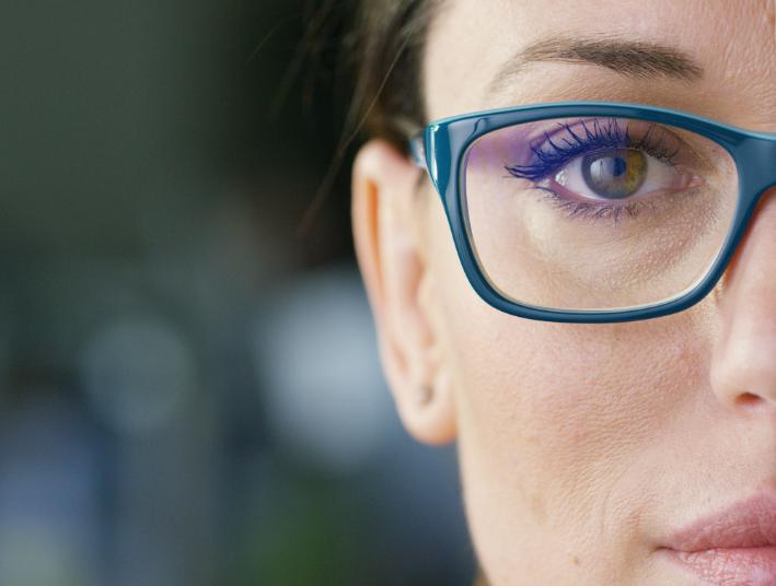
The Mind-Eye Institute gained worldwide attention for groundbreaking investigative and clinical work involving retinal processing. Indeed, expanding knowledge about the retina and application of 21st century optometric science enabled the Institute to achieve well documented, clinical successes in using specialized, therapeutic eyeglasses to directly alter brain activity and diminish fatigue, brain fog, concentration, memory difficulties, headaches, and other symptoms of traumatic brain injury, concussion, and stroke. These “brain” glasses have also proven effective in building undeveloped processing skills in children and adults with learning deficiencies, including those “on the spectrum,” namely autism and attention deficit hyperactivity disorder (ADHD).
By varying the amount, intensity, and angle of light passing through the retina, brain glasses help restore synchronization to patients’ disrupted sensory systems; alter their awareness, attention to, and understanding of what happens around them; restore visual processing skills; and bring a return of comfort and relief.
Remember, in today’s world of advanced science, help is available. TBI patients can find ways to manage fatigue, reconnect, and return quality to their lives. As former U.S. President Theodore Roosevelt once said, “Believe you can and you’re halfway there.”
Deborah Zelinsky, O.D., is a Chicago optometrist who founded the Mind-Eye Connection, now known as the Mind-Eye Institute. She is a clinician and brain researcher with a mission of building better brains by changing the concept of eye examinations into brain evaluations. For the past three decades, her research has been dedicated to interactions between the eyes and ears, bringing 21st-century research into optometry, thus bridging the gap between neuroscience and eye care.


